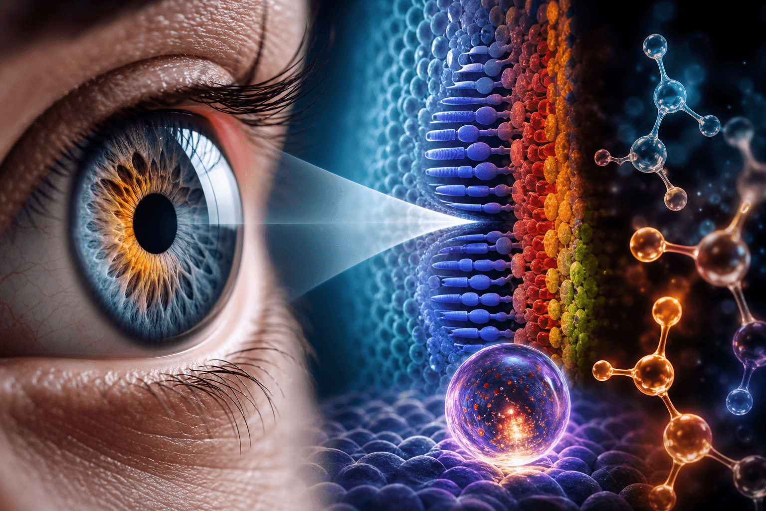
Scientists at Johns Hopkins University say they have identified a mechanism that helps build sharp central vision before birth, a finding that challenges a longstanding explanation of how the human retina develops. The study, published in the Proceedings of the National Academy of Sciences, used lab-grown retinal tissue to examine formation of the foveola, the small central part of the retina responsible for the sharpest vision. The researchers reported that vitamin A-related signaling and thyroid hormones work in sequence to shape the mix of cone cells in that region.
The work focused on cone photoreceptors, which support daytime and color vision and ultimately develop into blue, green or red cones. In the foveola, however, red and green cones dominate. The team said that during fetal development, a small number of blue cones appear in the developing foveola between weeks 10 and 12, but by week 14 those cells have changed into red and green cones. Researchers said this occurs through two mechanisms: the breakdown of retinoic acid, a molecule derived from vitamin A, reduces the formation of new blue cones, and thyroid hormones then drive remaining blue cones to convert into red and green cones.
The findings run against a model that has guided the field for decades, which held that blue cones formed in the retinal center and later moved outward. Robert J. Johnston Jr., the Johns Hopkins biologist who led the research, said the new data instead supports the view that the cells stay in place and change identity over time, though he added that the older theory cannot yet be fully ruled out. The study also highlights the difficulty of investigating this aspect of human vision in common animal models such as mice and fish, which do not develop the same photoreceptor arrangement.
Johnston and his colleagues say the discovery could eventually help improve retinal organoids and support future cell replacement therapies for diseases including macular degeneration and glaucoma. Johnston described the work as a step toward understanding the retinal center, which he said is among the first regions to fail in macular degeneration. Katarzyna Hussey, a co-author now at CiRC Biosciences, said the longer-term goal is to produce tailored photoreceptor cells that could be introduced into the eye to restore lost vision, while emphasizing that additional optimization and safety and efficacy studies would be needed before any clinical use.